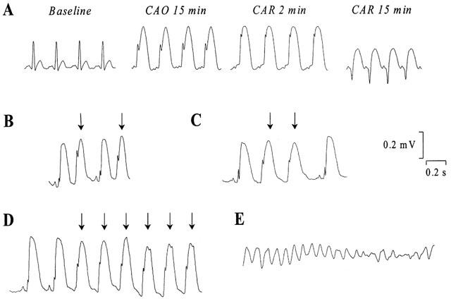
Figure 2.

Representative electrocardiogram recordings (A) at baseline, during coronary artery occlusion and reperfusion in a rabbit with normal rhythm, (B) of ventricular premature beats (indicated by the arrows), (C) of salvos of two ventricular premature beats, (D) of ventricular tachycardia and (E) of ventricular fibrillation. (CAO, coronary artery occlusion; CAR, coronary artery reperfusion).
