
Figure 3.
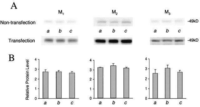
Expression of M1, M2, and M3 receptor subtypes in cultured cardiomyocytes. (A) Representative Western blots of M1, M2, and M3 in cardiomyocytes with or without transfection of muscarinic M1 – 3 receptors are shown. Different groups (n=3 in each group) of cultured cardiomyocytes were treated with control (a), 1 μM carbachol (b), and 1 μM MEG (c). (B) Blots of M1, M2, and M3 receptor proteins as shown in (A) with or without cDNA transfection were quantified with their internal standard cyclophilin A. Relative protein levels were presented as dividing transfection by non-transfection for each own receptor subtype. The expression efficiency was similar for these three muscarinic subtypes.