
Figure 2.
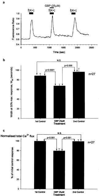
Gabapentin inhibited Ca2+ influx into cultured DRG as measured using fura-2 fluorescence imaging. (a) Trace from a single cultured DRG neurone showing a decrease in response duration and total Ca2+ flux by gabapentin (25 μM), but no significant change in the amplitude of the peak Ca2+ transient. (b and c) Bar charts showing the inhibitory actions of 25 μM gabapentin (GBP) on the duration of the Ca2+ transients evoked by 30 mM KCl measured at 50% of the peak amplitude (W50) and the total Ca2+ flux normalized with respect to the first control Ca2+ transient.