
Figure 5.
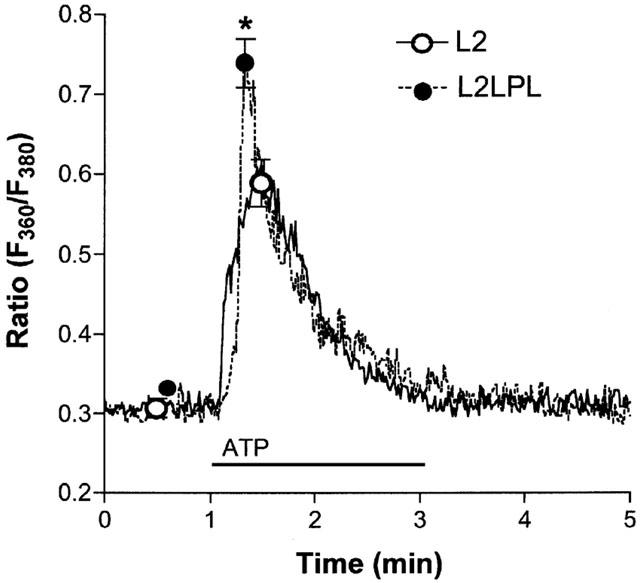
ATP-initiated Ca2+ signalling in single endothelial cells freshly isolated from aortae of L2 and L2LPL mice. Cells were isolated by enzymatic digestion, loaded with fura-2 and resuspended in KHS containing 2.5 mM CaCl2. In a microflorometer intracellular Ca2+ concentration in single endothelial cells was monitored as the ratio of 360 and 380 nm excitation at 510 nm emission (Ratio F360/F380). As indicated, 10 μM ATP was added to the superfusion. Tracings show representative experiments and points indicate the mean±s.e.mean (L2: n=8; L2LPL: n=16). *P<0.05 vs the effect of ATP in cells from L2 mice.