
Figure 7.
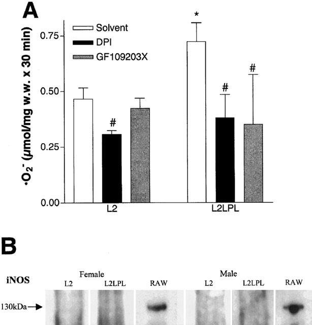
(A) Release of superoxide anions (•O2−) from aortae freshly isolated from L2 and L2LPL mice in the absence (solvent: 1% DMSO) or presence of 10 μM diphenylen iodonium (DPI) or after preincubation for 30 min with 1 μM GF109203X. Release of the •O2− was measured as the SOD-sensitive reduction of ferricytochrome c that was photometrically monitored at 550 nm. Columns represent the means±s.e.mean (L2: n=8; L2LPL: n=8). *P<0.05 vs results obtained in aortae of L2 animals and #P>0.05 vs the respective solvent control. (B) Western blot analysis on the expression of iNOS in aortae of female and male L2 and L2LPL mice. As a positive control, the cell lysate of the mouse macrophage cell line RAW 264.7 stimulated for 12 h IFNγ (10 ng ml−1) and LPS (1 μg ml−1) was used (BD Transduction Laboratories, Life Science Research, Vienna, Austria).