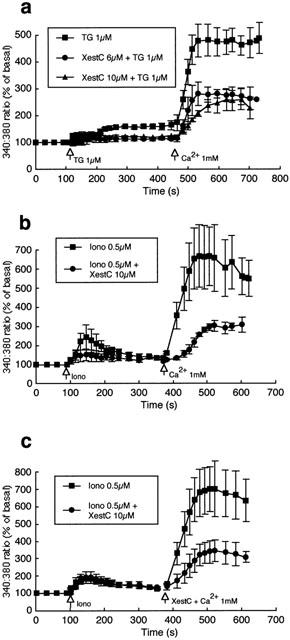
Figure 5.

Effects of xestospongin C on thapsigargin and ionomycin-induced Ca2+ entry in bovine endothelial cells. (a) Illustrates the inhibitory effect of xestospongin C (6 and 10 μM; n=5 and 4 respectively) compared to control (n=7) on capacitative Ca2+ entry induced by exposure of endothelial cells to thapsigargin (1 μM). (b) Shows the inhibitory effect of xestospongin C (10 μM) pretreatment on ionomycin (5 μM; n=3)-induced Ca2+ entry compared with control (n=3). (c) Illustrates that xestospongin C (10 μM) added at the time of Ca2+ addition to the superfusate also attenuates capacitative Ca2+ entry (n=4 for each condition). Results are presented as mean±s.e.mean. For clarity, representative error bars are shown.
