
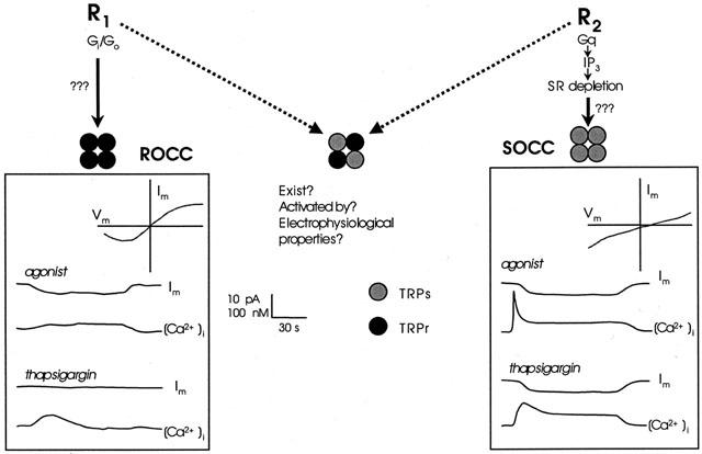
Figure 1.
Comparison of the responses mediated by ROCCs and SOCCs in smooth muscle. The left hand panel shows hypothetical changes in membrane current (Im) and intracellular calcium [Ca2+]i produced by a receptor agonist and the SERCA pump inhibitor thapsigargin in a cell possessing only ROCCs. The top right corner of the panel shows the current-voltage relationship for the receptor operated current. The functional channel is proposed to be formed from four ‘receptor coupled' (TRPCr) subunits (black circles). The right hand panel shows the equivalent responses but in this case from a cell possessing only SOCCs, formed from four ‘store-coupled' (TRPCs) subunits (grey circles). Note the difference in the current-voltage relationship and in the response to thapsigargin. A channel formed from a mixture of TRPs and TRPr subunits is depicted in the middle of the figure though firm evidence for the existence of such channels is lacking. See text for further details.