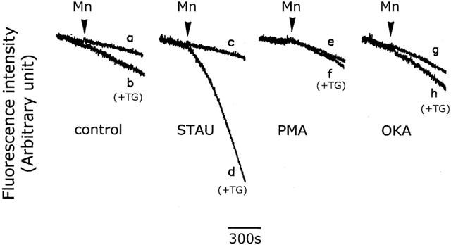
Figure 11.

Effect of protein phosphorylation on the thapsigargin-induced CCE. Fura-2-loaded cells were bathed in loading buffer containing 0.2 mM Ca2+ alone (control) (traces a and b), or plus 100 nM staurosporine (STAU) (traces c and d), 100 nM PMA (traces e and f), or 2 nM okadaic acid (OKA) (traces g and h). Five min later, 1 mM Mn2+ (traces a, c, e and g) or 1 mM Mn2+ plus 1 μM thapsigargin (TG) (traces b, d, f and h) was added, and the fluorescence quenching due to Mn2+ influx measured. The experiment was repeated six times using different batches of cells with similar results (n=15).
