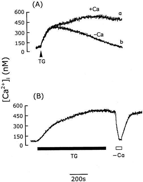
Figure 2.

Activation of CCE by thapsigargin in cerebellar astrocytes. (A) [Ca2+]i increases induced by 1 μM thapsigargin (TG) in the presence (trace a) or absence (trace b) of extracellular Ca2+. (B) After 10 min exposure to thapsigargin (1 μM) (TG), the [Ca2+]i remained elevated after removal of thapsigargin, but fell to the basal level when extracellular Ca2+ was removed and returned to the elevated level when Ca2+ was added back to the bathing buffer. Similar results were seen using five different batches of cells (n=15).
