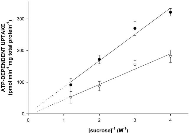
Figure 3.

Osmolarity dependence of N-methylquinidine transport into membrane vesicle preparations isolated from MDR1- or Mdr1b-overexpressing insect cells. Sf-MDR1 (closed circles) or Sf-Mdr1b (open triangles) membrane vesicles (25 μg of protein) were incubated for 25 s at 37°C sec in the presence of 8 μM [3H]-N-methylquinidine, and different concentrations of sucrose (0.25, 0.33, 0.5 and 0.8 M). Transport of [3H]-N-methylquinidine was determined as described in Methods. ATP-dependent uptake was calculated by subtracting values obtained in the presence of 4 mM AMP – PCP from those obtained in the presence of 4 mM ATP. Data points represent the mean±standard deviation of triplicate determinations.
