
Figure 4.
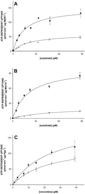
Uptake kinetics of N-methylquinidine, N-methylquinine, and vincristine by membrane vesicle preparations isolated from MDR1- or Mdr1b-overexpressing insect cells. Sf-MDR1 or Sf-Mdr1b membrane vesicles (25 μg of protein) were incubated in the presence of increasing concentrations of [3H]-labelled substrates. Initial uptake rates for N-methylquinidine, N-methylquinine, and vincristine were determined after an uptake period of 25, 50 and 15 s, respectively. Data presented are converted into uptake per min, and are corrected for Pgp-content of vesicle preparations. (A) Uptake of N-methylquinidine (closed circles) and N-methylquinine (open triangles) into Sf-MDR1 vesicles; (B) uptake of N-methylquinidine (closed circles) and N-methylquinine (open triangles) into Sf-Mdr1b vesicles; (C) uptake of vincristine into Sf-MDR1 (closed circles) and Sf-Mdr1b (open triangles) vesicles. Data points were analysed by non-linear curve fitting to an equation describing Michaelis – Menten kinetics using SigmaPlot. ATP-dependent uptake was calculated by subtracting values obtained in the presence of 4 mM AMP – PCP from those obtained in the presence of 4 mM ATP. Data points represent the normalized average uptake±standard error of the substrates into vesicles of three different membrane preparations (two for vincristine); the uptake into each vesicle preparation was determined three times and normalized for Pgp content. All individual data points were included in the non-linear curve fitting.