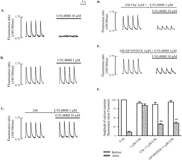
Figure 1.

Effects of U50,488H on electrically-stimulated [Ca2+]i transient in naive ventricular myocytes (A, B), and in myocytes pretreated with 1 μM U50,488H for 24 h in the absence of PKC inhibitors (C) or presence of 1 μM chelerythrine (D) or 1 μM GF109203X (E). [Ca2+]i transient was induced by electrical stimulation at 0.2 Hz. (A – E): Representative tracings. (F) Group results showing effects of 30 μM U50,488H transient in naive ventricular myocytes, and in myocytes pretreated with 1 μM U50,488H for 24 h in the presence of a PKC inhibitor. Values are mean±s.e. of 6 – 12 cells in 4 – 6 rats. All measurements were recorded at ∼15 min after administration of U50,488H. The amplitude of the [Ca2+]i transient without U50,488H is 100%. chelerythrine was added 60 min before addition of U50,488H (1 μM) for 24 h. One μM GF109203X was added 15 min before addition of U50,488H (1 μM). □num;□num;P<0.01 vs corresponding value in U50,488H group. Before: before addition of 30 μM U50,488H; after: after addition of 30 μM U50,488H. U50: U50,488H. Con: control. Che: chelerythrine.
