
Figure 6.
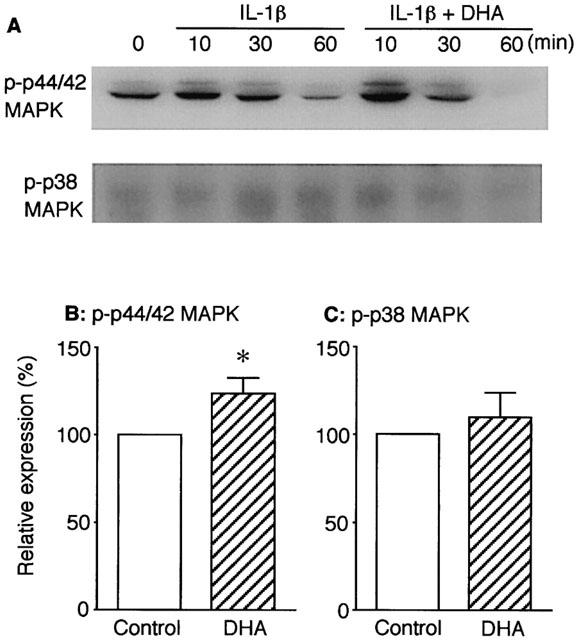
Effect of DHA on p44/42 MAPK and p38 MAPK activations induced by IL-1β in VSMCs. Expressions of phosphorylated p44/42 (p-p44/42) MAPK and p38 (p-p38) MAPK proteins were determined by Western blot analysis. (A) Representative Western blot images for p-p44/42 MAPK (upper) and p-p38 MAPK (lower). Cells were stimulated with 3 ng ml−1 IL-1β in the absence or the presence of DHA (30 μM) for the indicated time periods. (B and C) Summary of densitometric analysis for p-p44/42 MAPK and p-p38 MAPK, respectively, expressed as percentage taking each control (IL-1β alone) as 100%. Cells were stimulated with IL-1β in the absence or the presence of DHA for 10 min. Each column represents mean±s.e.mean of four separate experiments. *P<0.05 versus control.