
Figure 1.
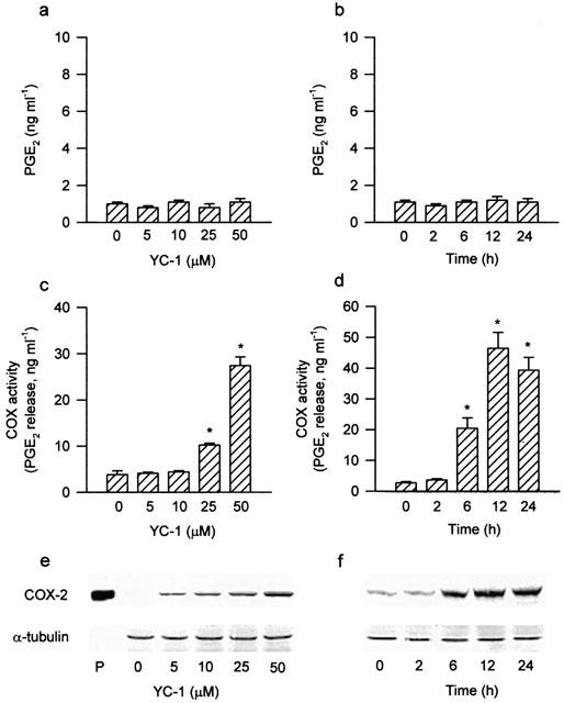
Effects of YC-1 on PGE2 release, COX activity and COX-2 expression in A549 cells. Treatment of the cells with various concentrations of YC-1 for 12 h (a) or YC-1 (50 μM) for the indicated time intervals (b) did not change the PGE2 release. YC-1 induced a concentration-dependent increase in COX activity (c) and COX-2 expression (e), and a time-dependent increase in COX activity (d) and COX-2 expression (f). The COX activity was measured by examining the PGE2 formation in the presence of 30 μM exogenous arachidonic acid for 30 min. Results are expressed as means±s.e.mean of four independent experiments performed in duplicate. *P<0.05 as compared with the basal level. In (e) and (f), cells were incubated with the indicated concentrations of YC-1 for 12 h (e) or YC-1 (50 μM) for various time intervals (f), and the extracted proteins were then immunodetected with COX-2 or α-tubulin specific antibody as described in Methods. The equal loading in each lane was demonstrated by the similar intensities of α-tubulin. Whole cell lysate of mouse macrophages (RAW 264.7) stimulated by LPS (1 μg ml−1) and INFγ (10 ng ml−1) for 12 h was used as a positive control (P).