
Figure 6.
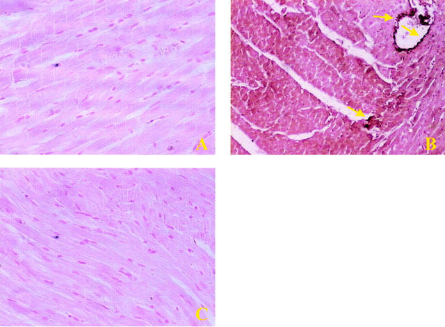
No positive staining for nitrotyrosine (A) was found in the left ventricular section from sham-operated rats. Sixty minutes after reperfusion immunohistochemical analysis for nitrotyrosine (B) show positive staining localized in the vascular wall (see arrows) and in the cardiomyocyte in the injured area. The intensity of the positive staining for nitrotyrosine (C) was significantly reduced in the left ventricular section from M40403-treated rats. Original magnification: ×150. Figure is representative of at least three experiments performed on different experimental days.