
Figure 9.
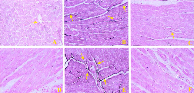
Staining of left ventricular section obtained from sham-operated rats with anti-ICAM-1 antibody showed a specific staining along vessels (arrow), demonstrating that ICAM-1 is constitutively expressed (A), no P-selectin staining was seen in sham animals. (D). Section obtained from IR shocked-rats showed intense positive staining (see arrows) for ICAM-1 (B) and for P-selectin (E) on vascular wall. The degree of endothelial staining for ICAM-1 (C) and for P-selectin (F) was markedly reduced in tissue section obtained from M40403-treated rats. Original magnification:×150. Figure is representative of at least three experiments performed on different experimental days.