
Figure 7.
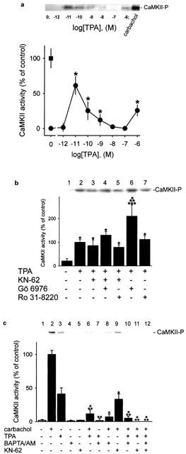
CaMKII is activated when resting parietal cells are treated with TPA. After treatment of parietal cells with TPA (0–1 μM), activated CaMKII (CaMKII-P) was detected in the membrane by immunoblot analysis (each upper panel). Bands of activated CaMKII were quantified by densitometry (each lower panel). Results are expressed as means±s.d. from 3–5 experiments. Absence of any chemical agent corresponds with basal acid secretion. (a) Resting parietal cells were treated with various concentrations of TPA (0–1 μM), and analysed for active CaMKII (black points). Controls were stimulated with carbachol (0.1 mM; black rectangle). *Significantly different from the corresponding control group (absence of any secretagogue or TPA), P<0.05. (b) Parietal cells were incubated with different protein kinase modulators (Gö 6976 (10 μM), KN-62 (20 μM), Ro 31-8220 (10 μM)) for 15 min with subsequent treatment by TPA (1 μM) for 30 min. *Significantly different from control (absence of TPA); **significantly different from TPA alone; ***significantly different from TPA+Gö 6976+KN-62, P<0.05. (c) Cells were pretreated with BAPTA/AM (0.1 mM) or KN-62 (20 μM) for 15 min, then incubated with carbachol (0.1 mM) or TPA (1 μM) or both for 30 min. *Significantly different from carbachol alone; **significantly different from TPA alone, P<0.01.