
Figure 1.
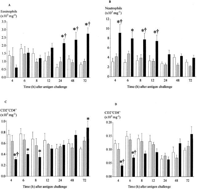
Time course of changes in the number of eosinophils (A), neutrophils (B), CD2+CD4+ (C) and CD2+CD8+ (D) cells recovered from lung tissue after antigen challenge in the Brown Norway rat. Antigen (ovalbumin)-sensitized Brown Norway rats were either unchallenged (open bars), exposed to inhaled saline (sham challenged, shaded bars) or challenged with inhaled antigen aerosol (solid bars). At a range of times after challenge, cells were isolated from lung tissue by enzymatic disaggregation. Cells were counted by light microscopy (a and b) or by flow cytometry (c and d). Group size was 10–12. Results represent mean±s.e.mean. *P<0.05 compared with unchallenged group; †P<0.05 compared with sham challenged group.