
Figure 2.
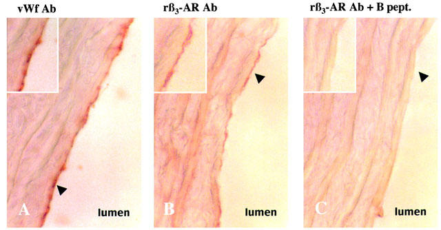
Comparison of von Willebrand factor (A) and β3-AR expression in rat thoracic aorta (B). Adjacent 10 μm thick sections were incubated with either von Willebrand antibody (vWf Ab; A) or rat β3-AR antibody (rβ3-AR Ab; B) revealed by peroxydase-conjugated second antiserum. Black arrowhead shows endothelium intensively stained with vWf and rβ3-AR Ab (A–B). Same staining after pre-absorption of rβ3-AR Ab with the blocking peptide (rβ3-AR Ab +B pept.; C). Micrographs X100.