
Figure 2.
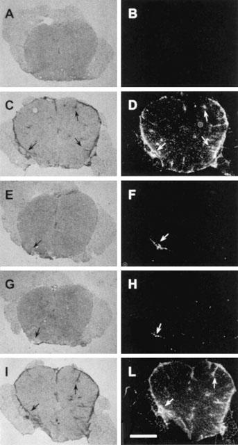
PHE and BZD but not BA reduce inflammatory infiltrates in the spinal cord of myelin-immunized rats. Inflammatory infiltrates in sections of the lumbar spinal cord of sham- (A, B) and myelin-immunized (C-L) rats at day 14 p.i. are shown with haematoxylin-eosin staining (A, C, E, G, I) or MHC II immunohistochemistry (B, D, F, H, L). Inflammatory infiltrates are not detectable in sham-immunized animals (A, B). Massive meningeal and perivascular inflammatory infiltrates (C) colocalize with MHC II+ cells (D) (arrows) in rats with EAE. Note the striking reduction of infiltrates in rats treated daily with 60 mg kg−1 PHE (E, F, arrows) or 200 mg kg−1 BZD (G, H, arrows). The treatment with 200 mg kg−1 BA does not affect immune infiltration (I, L, arrows). Each section is representative of one group of animals (n=5). Scale bar=550 μm.