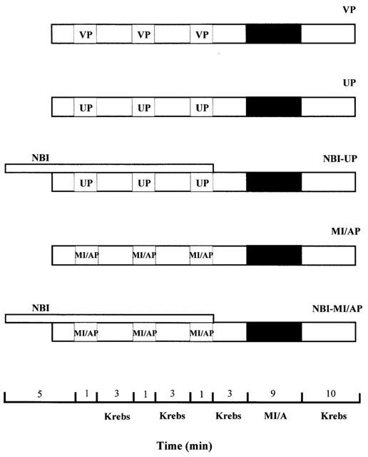
Figure 1.

Experimental design. After isolation, ventricular myocytes were allowed to stabilize for 30 min before treatment. Myocytes were then subjected to either pharmacological preconditioning with 30 μM U50 (UP) or metabolic inhibition/anoxia preconditioning (MI/AP) with 10 mM Na2S2O4+10 mM 2-DOG in glucose-free Krebs for three 1-min cycles separated by three min superfusion with normal Krebs solution. A group of cells were incubated with saline in Krebs as vehicle-preconditioning control (VP). Another two groups of myocytes were subjected to UP or MI/AP in the presence of 5 μM nor-BNI, which was administered 5 min before and throughout the preconditioning period. These two groups were NBI-UP and NBI-MI/AP, respectively. Then the ventricular myocytes in all groups were subjected to MI/A with 10 mM Na2S2O4+10 mM 2-DOG in glucose-free Krebs solution for 9 min followed by superfusion with normal Krebs solution for 10 min–reperfusion.
