
Figure 4.
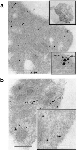
Immunogold electron microscopy examination of TIMP-4, MMP-2 and MMP-9 in non-aggregated platelets. Large gold particles (15 nm) label TIMP-4, while small particles (5 nm) label MMP-2 (A) or MMP-9 (B). TIMP-4 and MMP-2 co-localize as shown by arrows (A, lower inset). No IgG-related immunoreactivity was detected in control experiment (A, upper inset). Co-localization of TIMP-4 and MMP-9 is less apparent. Bar: 0.5 μm, inset bar: 0.1 μm.