
Figure 4.
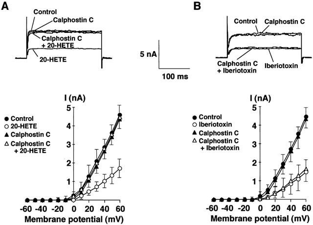
Effects of calphostin C on whole-cell K+ current inhibited by 20-HETE and iberiotoxin. (A) Effect on 20-HETE-induced inhibition of whole-cell K+ current. (B) Effect on iberiotoxin-induced inhibition of whole-cell K+ current. Upper panel, representative tracings of outward whole-cell K+ current elicited by depolarizing pulses to +60 mV from a holding potential of −70 mV in the absence (in A and B) or presence of 100 nM 20-HETE (in A), 1 μM calphostin C (in A and B), 1 μM calphostin C plus 100 nM 20-HETE (in A) or 1 μM calphostin C plus 100 nM iberiotoxin (in B). Lower panel, current-voltage relationships of whole-cell K+ current for the same cell shown in the upper panel in the absence and presence of 100 nM 20-HETE (in A), 100 nM iberiotoxin (in B), 1 μM calphostin C (in A and B), 1 μM calphostin C plus 100 nM 20-HETE (in A) or 1 μM calphostin C plus 100 nM iberiotoxin (in B). Whole-cell K+ current was activated by incremental 10 mV depolarizing steps from a holding potential of −70 mV to +60 mV. Each point represents the mean±s.e.mean of five experiments.