
Figure 4.
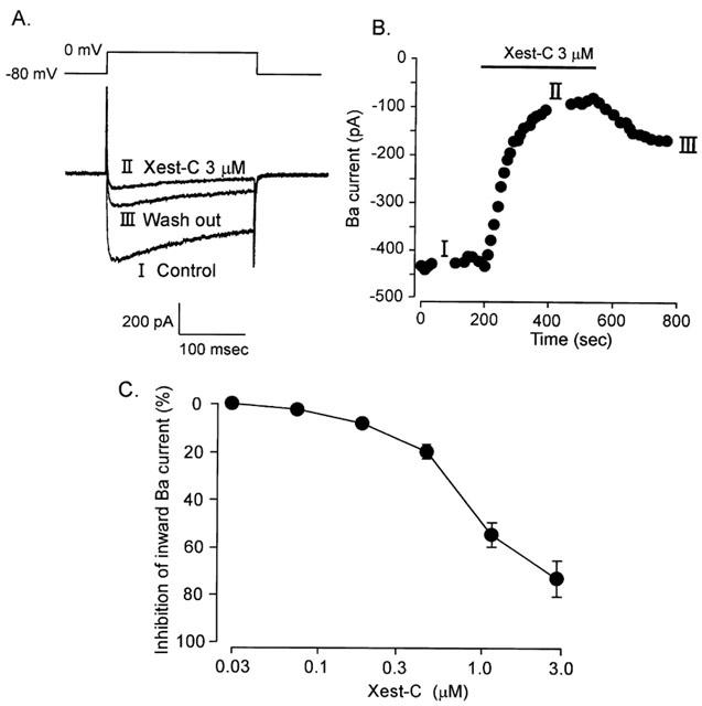
Effects of xestospongin-C on IBa through Ca2+ channels in single guinea-pig ileal smooth muscle cells. IBa was elicited by 80 ms depolarisation from −60 to 0 mV at 0.1 Hz. (A) Typical tracings of IBa in the presence or absence of 3 μM xestospongin-C. (B) Changes in peak amplitude of IBa with time. Xestospongin-C (3 μM) was applied during the period indicated by a horizontal bar. Time 0 min indicates the start of IBa recording under conditions in which the K+ current was blocked by internal diffusion of Cs from the recording pipette for about 3 min after the patch membrane was ruptured. (C) Effects of various concentrations of xestospongin-C on IBa (n=4–6).