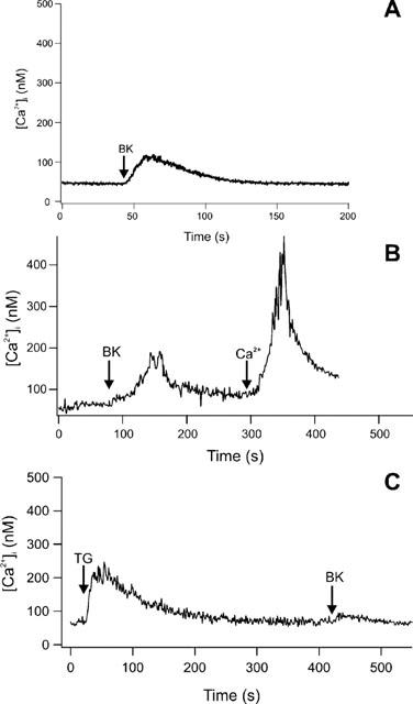
Figure 4.

(A) Increase of [Ca2+]i in peritubular cells by stimulation with BK at a final concentration of 10 nM in a nominally Ca2+-free external HBSS. The stimulus was followed by a diminished calcium response compared to the stimulation in the presence of external calcium. (B) Addition of Ca2+ (2 mM) after BK stimulation in nominally Ca2+-free external HBSS led to an instantaneous and linear rise of [Ca2+]i (C) The SERCA-pumps of the endoplasmic reticulum were blocked by 5 μM thapsigargin. After a transient rise in [Ca2+]i cells were stimulated with BK (10 nM). No substantial rise in [Ca2+]i was detectable. All figures show one typical experiment out of at least seven.
