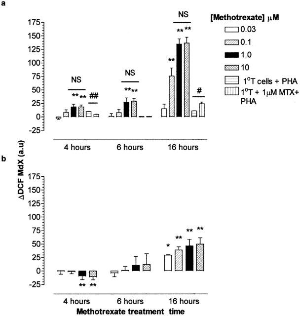
Figure 4.

Methotrexate mediated alterations in the cytosolic peroxide levels of Jurkat T-cells, primary T cells and U937 monocytes. Determination of the kinetics for the oxidation of DCFH to DCF. Jurkat T-cells or primary T cells (a) or U937 monocytes (b; 2×10⁶ ml−1) was undertaken in cells which were serum starved in RMPI 1640 for 4 h prior to the addition of 0–100 μM methotrexate for 4, 6 or 16 h. Cells were treated with 50 μM DCFH-DA as described in Methods. At the end of the treatment period, cell samples were analysed immediately for DCF fluorescence by flow cytometry. The median X (MdX) DCF fluorescence of 10,000 cells was analysed per sample. ΔDCF represents the difference in MdX DCF of methotrexate treated cells from that of vehicle treated cells for each time point. All incubations were performed at 37°C in a humidified, 95% air, 5% CO2 atmosphere. The data is expressed as the mean±s.e.mean of four individual experiments where *(P<0.05) and **(P<0.01) were considered significantly different from control samples by one-way ANOVA followed by Tukey's post hoc test analysis. a.u., arbitrary units; NS, no significant difference.
