Figure 1.

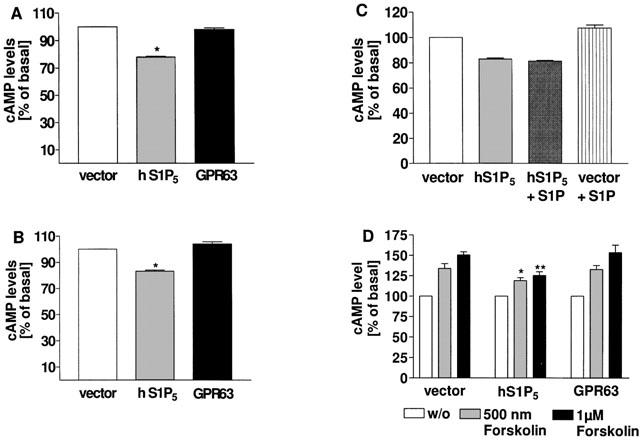
cAMP accumulation in HEK 293 cells stably expressing the human S1P5 receptor, GPR63 or vector. Cells stably expressing the indicated receptors or pcDNA3.1(+) vector DNA as a control were grown in medium containing 10% FBS (A) or charcoal-stripped FBS (csFBS) (B) for 24 h and cAMP levels quantitated in the presence of 500 μM IBMX with the alpha-screen® kit as described in Methods. (C) Transfected cells were cultivated in csFBS for 24 h and stimulated with 1 μM S1P for 30 min prior to quantification of cAMP levels. (D) Transfected cells were stimulated with forskolin for 30 min in the presence of 500 μM IBMX and cAMP levels determined as described above. In cells expressing the human S1P5 receptor basal cAMP levels were significantly lower than in vector or GPR63 expressing cells, whether they were cultivated in the presence (A) or absence (B) of lipids: *P<0.05 indicates significant difference versus cAMP levels in vector transfected cells. Addition of S1P did not significantly affect intrinsic S1P5 receptor-mediated cAMP inhibition (C). Forskolin stimulation of S1P5 receptor transfected cells resulted in significant lower cAMP levels as compared with vector or GPR63 transfectants (D): *P<0.05 and **P<0.01 indicate significant differences versus cAMP levels in vector transfected cells under corresponding conditions. Data are means±s.e.mean (n=4–10).
