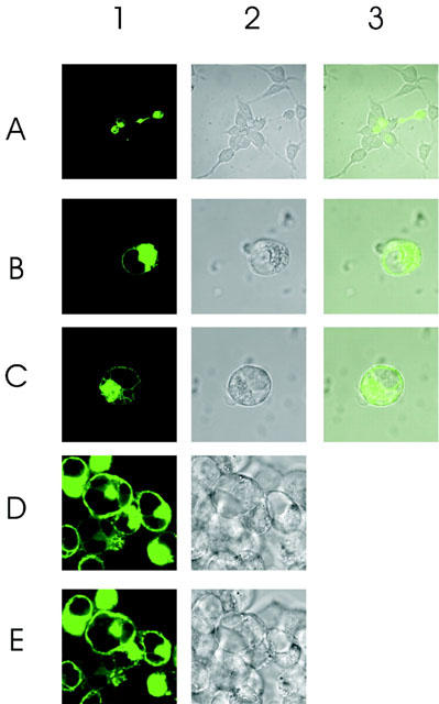
Figure 7.

Cellular distribution pattern of GFP-tagged wild type S1P5 receptor. (A) C-terminally GFP-tagged S1P5 receptor was expressed transiently in HEK293 cells and images were analysed confocally. A1–3: the majority of transfected cells are round whereas non transfected cells display the characteristic HEK293 cell morphology (A2,3). B1–3: images frequently observed showing a significant amount of GFP fluorescence in intracellular compartments. Some cells (C1–3) display GFP fluorescence at the plasma membrane. D: cells were stimulated with 10 μM S1P for 10 min. E: cells were stimulated with 10 μM S1P for 20 min. Cell rounding does not allow evaluation of internalization responses.
