
Figure 5.
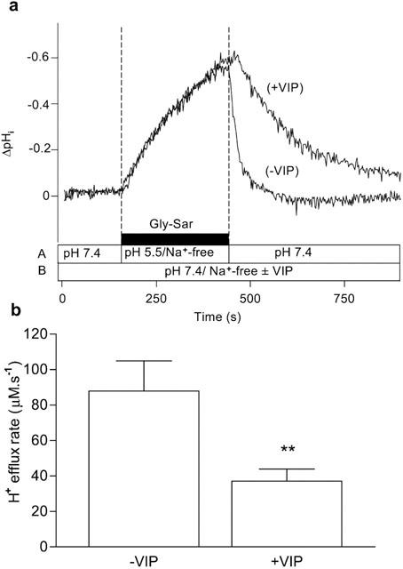
Intracellular pH recovery from a Gly-Sar-induced acidification in the presence and absence of VIP. Caco-2 cell monolayers were exposed to Gly-Sar (20 mM) in the apical superfusate (apical pH 5.5, Na+-free solution) for 5 min (the basolateral membrane was superfused with a Na+-free Krebs' solution (pH 7.4) throughout the experiment). The apical superfusate was then replaced with a pH 7.4, Na+- containing, Gly-Sar-free solution and pHi recovered to basal levels. This ‘control' manoeuvre was performed in the absence of VIP (−VIP). After the ‘control' (−VIP) manoeuvre was completed the cell monolayer was exposed to VIP (5 nM) for 10 min at the basolateral membrane before the manoeuvre (acidification for 5 min with Gly-Sar followed by recovery at pH 7.4/Na+) was repeated in the presence of 5 nM basolateral VIP (+VIP) (total exposure time to VIP before recovery was 15 min). (a) Top panel: The trace represents the composite response seen in a single monolayer to successive recoveries in the absence (−VIP) and presence (+VIP) of VIP. The composition and pH of the apical (A) and basolateral (B) solutions are indicated by the horizontal bars. The results are expressed as a ΔpHi. (b) Bottom panel: The H+-efflux rate was calculated in each individual monolayer from the rate of the initial 30 s of recovery (after removal from the apical chamber of the Gly-Sar/pH 5.5/Na+-free solution) both in the absence (−VIP) and presence (+VIP) of VIP (as described in Methods). Data are mean±s.e.mean (n=8). **, P<0.01.