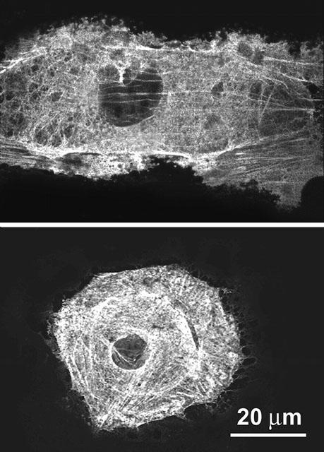
Figure 9.

Representative confocal micrographs of cultured rat aortic smooth muscle cells immunostained to reveal α-smooth muscle actin in resting conditions and upon addition of 10 nM LTD4. In the resting cell, cytoplasmic actin is in the form of typical stress fibres, whereas in the LTD4-stimulated cell actin is distributed evenly throughout the cytoplasm and the overall cell shape suggests cell contraction. Cells were fixed 10 min after addition of the stimulus (see Methods). Immune reaction was revealed with Alexa 488 nm-labelled goat anti-mouse antibodies.
