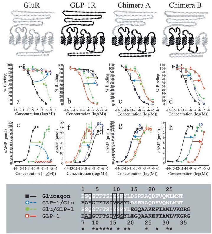
Figure 1.

Competition binding and functional analyses of chimeric glucagon/GLP-1 peptides and chimeric glucagon/GLP-1 receptors. The upper panel illustrates the glucagon receptor (GluR) in grey, the GLP-1 receptor (GLP-1R) in black and two chimeric receptors composed of elements of the glucagon and GLP-1 receptor. Below each receptor are the corresponding binding and functional analyses. Each figure is representative of three or more independent experiments performed in triplicates (binding analyses) or duplicates (functional analyses). Competition binding analyses of the glucagon receptor and chimera A and B were performed using 125I-glucagon as the tracer. Competition binding analysis of the GLP-1 receptor was performed using 125I-GLP-1 as the tracer. (a) and (e): Competition-binding analysis (a) and functional analysis (e) of the glucagon receptor. (b) and (f): Competition-binding analysis (b) and functional analysis (f) of the GLP-1 receptor. (c) and (g): Competition-binding analysis (c) and functional analysis (g) of chimera A. (d) and (h): Competition-binding analysis (d) and functional analysis (h) of chimera B. The lower panel shows a sequence alignment of glucagon, GLP-1 and the two chimeric peptides GLP-1/Glu and Glu/GLP-1. The glucagon sequence is shown in white, the GLP-1 sequence is shown in black and a star illustrates amino acid identity. Glucagon is numbered 1–29 and GLP-1 is numbered 7–37.
