
Figure 7.
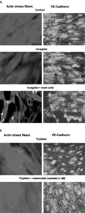
Actin stress fibres increase, while VE-cadherin immunoreactivity decreases after exposure of cultured HPAECs to ioxaglate (100 mg iodine ml−1), ioxaglate+mast cells, and tryptase (7×10⁻³ units ml−1). The increase in actin stress fibres and the decrease in VE-cadherin induced by ioxaglate+mast cells or tryptase are blocked by nafamostat mesilate (1 nM).