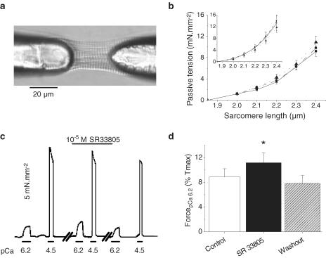
Figure 3.
Effects of SR33805 on passive and active forces in skinned cardiomyocytes. (a) Picture of a rat-skinned ventricular myocyte glued to glass needles and held at 1.9 μm SL. (b) Passive tension–SL relations established in the relaxing solution (pCa 9) by stretching the cells stepwise (in control solution (solid line) and during the successive applications of 10−7 (dashed line) and 10−5 M (dotted line) SR33805 (n=8)). Inset: stretching was induced by a controlled ramp at 0.1 length s−1 (n=8). (c) Original recordings of the tension elicited by a skinned cell maintained at 2.3 μm SL and submitted to stepwise superfusion of submaximal (pCa 6.2) and maximal (pCa 4.5) Ca2+-ativating solutions in the absence and in the presence of 10−5 M SR33805. The third series of recording obtained 3 min after washout demonstrates the recovery of the SR33805 effects. (d) Averaged contractile forces elicited at pCa 6.2 relative to maximal Ca2+ -activated forces in control conditions and in the presence of 10−5 M SR33805 on cells held at 2.3 μm SL (n=6). Washout was estimated at 3–5 min. *P<0.05.

