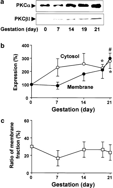
Figure 2.
Immunoblotting analysis of cPKCs from nonpregnant and pregnant myometria. Uterine fractions were prepared as described in ‘Methods'. (a) Upper and lower panels show representative expression of PKCα in the membrane fraction and PKCβI in the cytosolic fraction, respectively. (b) Statistical results in the cytosolic and membrane fractions were obtained from three independent experiments. The level of PKCα in nonpregnant myometrium was defined as 100%. (c) Quantification of the gestation-induced changes in the membrane distribution of PKCα. The ratio of membrane fractions was expressed as a percentage of the total expression refers to the sum of immunoreactivity in the cytosolic and membrane fractions. *, # Denote significant differences from the results of the cytosolic and the membrane fractions in nonpregnant myometrium, respectively (P<0.05).

