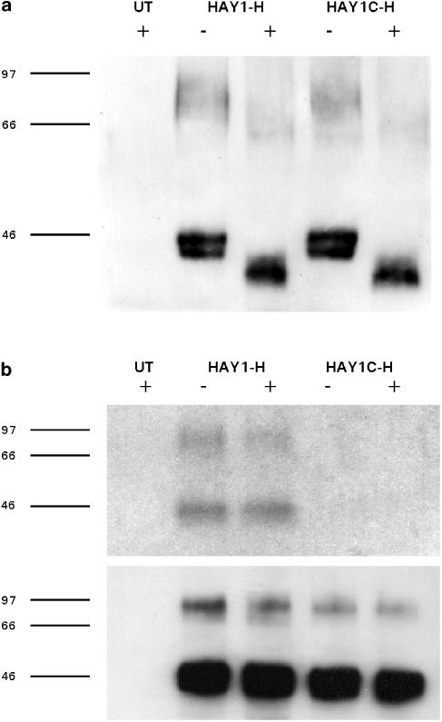
Figure 1.
(a) Anti-HA immunoprecipitates from untransfected CHO cells (UT), HAY1-H or HAY1C-H clones were resolved by 7.5% SDS–PAGE with (+) or without (−) prior treatment with PNGase F and transferred to a polyvinyl difluoride membrane. The blot illustrated (representative of four experiments) was probed with the primary antibody CT/2, raised against a Y1 receptor C terminal peptide. (b) Cells were labelled with [3H]palmitic acid and control (−) or 300 nM NPY-treated (+) samples were immunoprecipitated with directly conjugated anti-HA agarose. After resolution by 10% SDS–PAGE, gels were subjected to fluorography to detect [3H] incorporation (upper panel, one of two experiments). The lower panel illustrates the equivalent portion of a Western blot performed in parallel to confirm equal loading of HAY1-H and HAY1C-H receptors.

