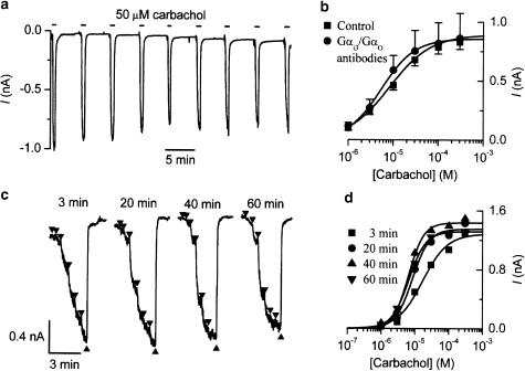
Figure 4.
The depression of mIcat by Gαi3/Gαo antibodies is prevented by carbachol application shortly after breakthrough to the whole-cell recording mode. (a) Current trace from a cell dialysed with anti-Gαi3/Gαo antibodies (Calbiochem; 1 : 200 v v−1) and repeatedly exposed to carbachol (50 μM) every 5 min starting 90 s after breakthrough. Note that the decline in mIcat was no more than the usual desensitization seen in control cells. (b) Averaged concentration–effect curves for carbachol-activated mIcat in cells dialysed with (n=4) or without (same data as shown in Figure 3a; n=53) the Gαi3/Gαo antibody. The former cells were exposed to carbachol (50 μM) for about 1 min at 1–3 min after the breakthrough. See text for details. (c) Current traces from a cell dialysed with Gαi3/Gαo antibodies and exposed to ascending carbachol concentrations (1–300 μM, shown by triangles) applied every 20 min starting 3 min after the breakthrough. (d) Concentration–response curves for the experiment shown in (c). Note that no inhibition of the agonist curve by Gαi3/Gαo antibody occurred using this protocol even at 60 min.

