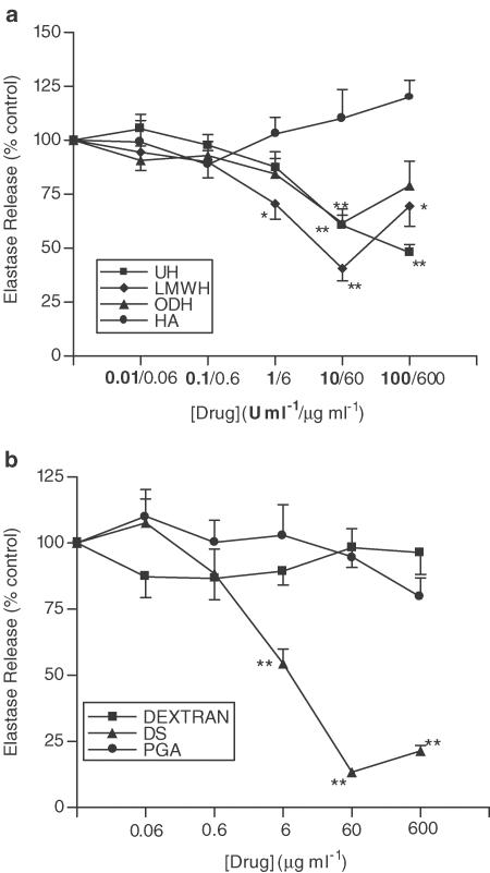
Figure 7.
Effects of UH, ODH, LMWH, HA (a), DS, dextran and PGA (b) upon PAF-induced elastase release in the presence of TNF-α. Neutrophils were placed into microfuge tubes at a concentration of 2.5 × 10⁶ cells ml−1, in the presence of 5 μg ml−1 cytochalasin B and the presence or absence of 100 U ml−1 TNF-α and inhibitor. After 30 min, 10−6 M PAF was added to the cells for 45 min, and cells were then centrifuged, samples of supernatant taken and incubated for 1 h with elastase substrate. After 1 h at room temperature, the coloured product was measured colorimetrically at 405 nm, using a microplate reader. Results from experiments on cells from at least six donors, each performed in duplicate, are expressed as mean ± s.e.m. values, expressed as a percentage of control values from experiments carried out without drug (*P<0.05 and **P<0.01, when compared with media control).

