Figure 1.
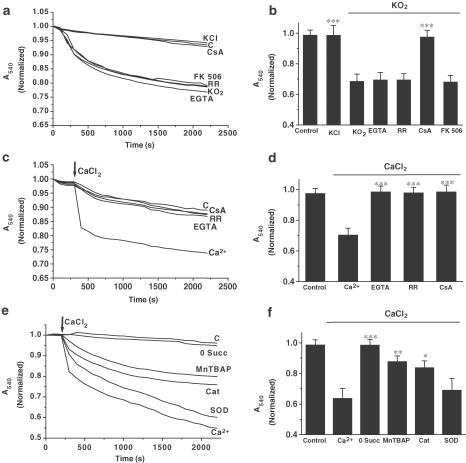
KO2 and high Ca2+ induce mitochondrial swelling. A–B. KO2 addition (5 μmol mg−1 protein; arrow), induces mitochondrial swelling in brain-isolated mitochondria in an MPTP sensitive manner, where Ca2+ cycling is not involved. Mitochondrial suspensions were incubated during 15 min with CsA (10 μM), FK 506 (200 nM), Ruthenium Red (RR; 5 μM), or EGTA (1 mM), prior to addition of KO2. Control (c) and the effect of KCl (5 mM) addition on non-treated mitochondria are also shown. C–D. Calcium addition (15 μmol mg−1 mitochondria) induces mitochondrial swelling in brain-isolated mitochondria. Mitochondrial suspensions were incubated during 15 min with CsA (10 μM), Ruthenium Red (RR;5 μM), or EGTA (1 mM) before Ca2+ addition. Control non-treated mitochondria (c) are also shown. E–F. Ca2+-induced swelling is ROS and succinate dependent. In absence of succinate (0 Succ), Ca2+ failed to induce mitochondrial swelling. Mitochondria were preincubated for 15 min with MnTBAP (10 nM), catalase (Cat;1750 U) or SOD (450 U), prior to Ca2+ addition (15 μmol mg−1 mit). Control nontreated mitochondria (c) are also shown. Lines represent mean values of one experiment performed by triplicate. Histograms (panels b, d, f) represent mean values±s.e.m. of normalized A540 at 2200 s from at least five different mitochondrial preparations. ***P<0.001;**P<0.01;*P<0.05 as compared with KO2 or Ca2+-treated mitochondria in absence of the drug.

