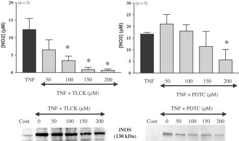
Figure 2.
TNFα-induced iNOS expression and involvement of NF-κB in differentiated 3T3-F442A adipocytes. Differentiated 3T3-F442A adipocytes were treated or not for 20 h with TNFα in the presence of increasing concentrations of two NF-κB inhibitors, TLCK (50–200 μM) or PDTC (50–200 μM). Nitrites were assessed in cell supernatants. Results are expressed as mean±s.e.m. from three independent experiments performed in duplicate. * indicates a statistically significant difference versus TNFα-treated cells (one-way ANOVA followed by Dunnett's post hoc test, P<0.05). iNOS protein was detected by Western blot. A representative autoradiography from three independent experiments performed in duplicate is shown.

