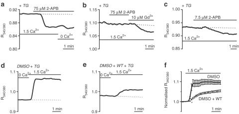
Figure 5.
2-APB and WT inhibit cerebral arteriolar SOCs. Arterioles were in standard (1.5 mM Ca2+) or Ca2+-free (0.4 mM EGTA) bath solution. All solutions contained 10 μM D600. (a–c) Effects of 2-APB in TG-pretreated arterioles. (a) 2-APB (75 μM) rapidly reduced [Ca2+]i. (b) In another arteriole there was no effect of 75 μM 2-APB but Gd3+ reduced [Ca2+]i, confirming SOC activation. (c) 2-APB (7.5 μM) slowly reduced [Ca2+]i. (d–f) Preincubation with WT inhibited Ca2+ entry in TG-pretreated arterioles. Arterioles were first incubated with either 0.1% DMSO (d) or 0.1% DMSO and 10 μM WT (e) in standard (1.5 mM Ca2+) bath solution. After this period, arterioles were incubated with TG in Ca2+-free (0.4 mM EGTA) bath solution. The increase in [Ca2+]i associated with the readdition of 1.5 mM Ca2+ was reduced in WT-pretreated arterioles. (f) Mean±s.e.m. data for experiments as shown in (d) and (e), normalised to the value of R340/380 at the point of addition of 1.5 mM Ca2+. DMSO group, n/N=18/4. WT group, n/N=20/4. P<0.001 for the data point marked * and all subsequent data points.

