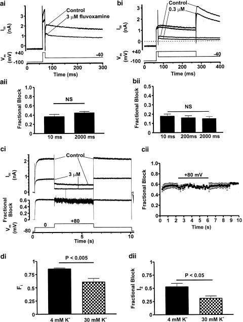
Figure 5.
State-dependence of block. Panel (a) shows representative IHERG traces recorded prior to and the first record following equilibration in 3 μM fluvoxamine, during which period membrane potential was held at −100 mV to maintain all channels in the closed state. Membrane potential was then briefly stepped to +40 mV for 10 ms prior to repolarisation to −40 mV to evoke IHERG tails. Fractional block of IHERG tails is summarised in panel (aii) (10 ms, n=4) and for comparison is shown alongside fractional block data taken from the concentration–response (unpaired t-test, P>0.15, NS denotes non-significance). Panel (b) To further investigate closed vs rapid open-state block, fluvoxamine concentration was reduced by 10-fold in order to reduce the rate of drug-channel association to the open state. Experiments were performed as in (ai) with the addition of a further trace in which membrane potential was stepped to +40 mV for 200 ms to investigate time-dependent changes in block. Data are summarised in panel (bii) showing fractional block following 10 and 200 ms activating steps. For comparison, fractional block data for 0.3 μM fluvoxamine is shown from the concentration–response relation. (ANOVA, P>0.6; NS denotes non-significance). Data from panels (a) and (b) are concordant with a component of closed state, although a component of very rapid open-state block cannot be excluded. Panel (c) shows a protocol similar to that described in Figure 4a with an additional 4 s step to +80 mV to produce profound IHERG inactivation. Representative IHERG traces are shown in the absence and presence of 3 μM fluvoxamine; plotted below is fractional block for that cell. Mean fractional block data are shown in 100 ms intervals in (cii) (n=4 cells). Bar indicates step to +80 mV. No significant change in block was observed following profound inactivation (ANOVA, P>0.9), suggesting that fluvoxamine's interaction with HERG is not hindered by strong inactivation. Panel (di) shows summarised data from experiments in which the fraction of inactive channels (Fi) following a 150 ms activating step from −80 to 0 mV was altered by elevating [K+]o from 4 to 30 mM (n=5 and 4, respectively, unpaired t-test) (see ‘sampling protocol' ‘Methods'; this time point was chosen as by 150 ms Fi had reached steady-state). Panel (dii) summarises the effect of modulating Fi on fractional block of the inactivation relieved current (I2, see ‘Methods') using an identical ‘sampling protocol' with an activating step to 0 mV for 150 ms. Reducing Fi by elevating [K+]o resulted in a significant reduction in fractional block of IHERG by 3 μM fluvoxamine (unpaired t-test, P<0.05).

