Figure 3.
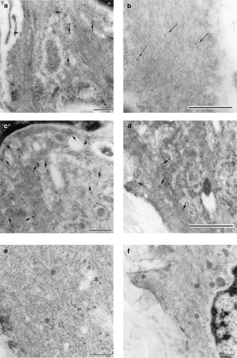
Transmission electron micrograph of ultrathin cryosections from the muscle layer of the foetal mouse ductus arteriosus. (a) Immunogold labelling for COX1; note the gold particles being clustered in the cytoplasm (indicated with arrows) which surrounds the nucleus (N) and in the plasma membrane (arrowheads). (b) Immunogold labelling for COX1 in the peripheral cytoplasm; label (arrows) is not as dense as that seen in the perinuclear cytoplasm. (c) Immunogold labelling for COX2 in the perinuclear cytoplasm; note clusters of label in proximity of the nucleus (N). (d) Immunogold labelling for COX2; note few clusters of label (arrows) in the peripheral cytoplasm and along the plasma membrane. (e) Immunogold labelling for cytosolic PGE synthase; label was scattered throughout the cytoplasm (asterisks). (f) Immunogold labelling for microsomal PGE synthase; label was found diffusely throughout the perinuclear (N) and peripheral cytoplasm (asterisks). Bar represents 0.25 μm (panels a, c, e, f) or 0.5 μm (panels b and d).

