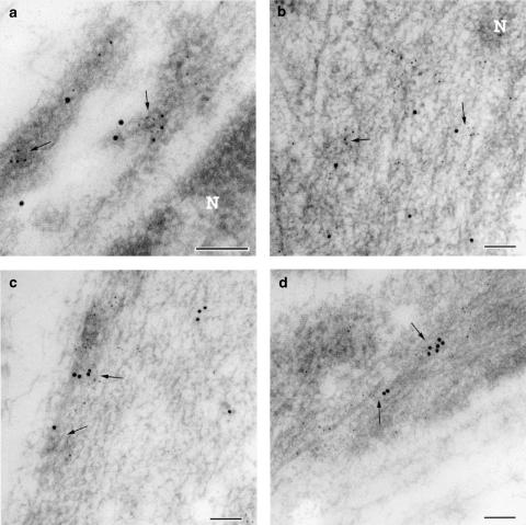
Figure 4.
Transmission electron micrograph of ultrathin cryosections from the muscle layer of the foetal mouse ductus arteriosus. (a) Dual immunogold labelling for COX1 (particle, 10 nm) and microsomal PGE synthase (particle, 5 nm); note colocalisation of the two labels (arrows) in vesicle-like structures close to the nucleus (N). (b) Dual immunogold labelling for COX2 (particle, 10 nm) and microsomal PGE synthase (particle, 5 nm); colocalisation of the two labels (arrows) in proximity of the nucleus (N). (c) Dual immunogold labelling for COX1 (particle, 10 nm) and microsomal PGE synthase (particle, 5 nm) in the peripheral cytoplasm; colocalisation of the two labels in vesicle-like structures (arrows). (d) Dual immunogold labelling for COX2 (particle, 10 nm) and microsomal PGE synthase (particle, 5 nm) in the peripheral cytoplasm; colocalisation of the two labels (arrows). Note that in both perinuclear and peripheral cytoplasm, COX2 colocalised more frequently than COX1 with microsomal PGE synthase. Bar represents 0.1 μm.

