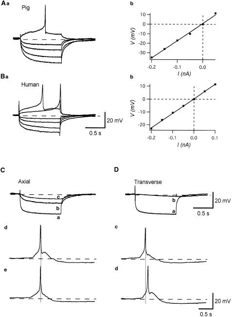
Figure 3.
Passive membrane properties of detrusor smooth muscle of the human and pig bladder. Overlaid traces show the membrane potential changes in the pig (Aa) and human (Ba) bladder induced by intracellular current injection of +0.1, +0.05, −0.05, −0.1, −0.15 and −0.2 nA for 1 s. The relation between the amplitude of injected currents and resultant membrane potential changes was linear in both cases. The input resistance calculated from the slope of I–V relation for pig detrusor was 160 MΩ (Ab), and for human detrusor, 110 MΩ (Bb). Resting membrane potentials were −40 mV in (A) and −39 mV in (B). Intracellular injection of inward current of −0.2 nA for 2 s induced a hyperpolarization with an amplitude of some 25 mV (Ca). The electrotonic potential recorded from a cell located about 200 μM apart in the axial direction had an amplitude of some 13 mV (Cb) and that in a cell located 400 μM apart axially had an amplitude of some 7 mV (Cc). In the same preparation, an injection of inward current of −0.2 nA for 2 s caused a hyperpolarization with an amplitude of some 25 mV (Da). The resultant membrane potential changes in a cell located 50 μM away in the transverse direction had an amplitude of some 4 mV (Db). In a different preparation, when two electrodes were placed 400 μM apart axially, action potentials recorded from both electrodes occurred almost simultaneously (Cd, e). When electrodes were placed 100 μM apart transversely, some 100 ms of delay between the peaks of paired action potentials could be detected (Dc, d). Resting membrane potential was −40 mV in (C) and (D) and −43 mV in (E) and (F).

