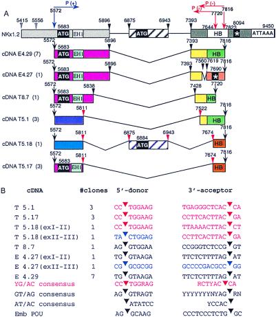
Figure 3.
(A) Nkx-1.2 gene structure, intron-exon junctions, and schematic representation of deduced Nkx-1.2 proteins. (B) Nucleotide sequences at the 5′ donor and 3′ acceptor splice sites of six species of Nkx-1.2 cDNA. Nkx-1.2 cDNA species cloned from embryo mRNA or testis mRNA are aligned to genomic Nkx-1.2 DNA. The first letter of each clone name (E or T) means that the cDNA clone was derived from embryo or adult testis poly(A)+ RNA, respectively. The number of clones isolated and sequenced for each species of cDNA is shown enclosed by parentheses. Noncanonical splice sites are indicated by red arrowheads; canonical splice sites are shown as black arrowheads. Horizontal arrows indicate the position of the primers used for the RT-PCR experiment. A star represents a termination codon.

