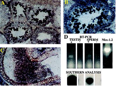
Figure 5.
Nkx-1.2 gene expression in germ cells. (A–C) In situ hybridization of Nkx-1.2 mRNA in the seminiferous epithelium of the testis (A and B) and in the epididymal duct of the adult mouse (C). (D) RT-PCR amplification of Nkx-1.2 cDNA from testis and spermatozoa RNA. In situ hybridization was performed as described in the text. PCR amplification was performed with primers +P1 and −P1 shown in Fig. 1. PCR products were separated by size by agarose gel electrophoresis, and the DNA bands were transferred to nylon membranes. Southern analysis was performed by hybridization with a 32P-labeled oligodeoxynucleotide probe corresponding to primer −P2 shown in Fig. 1.

