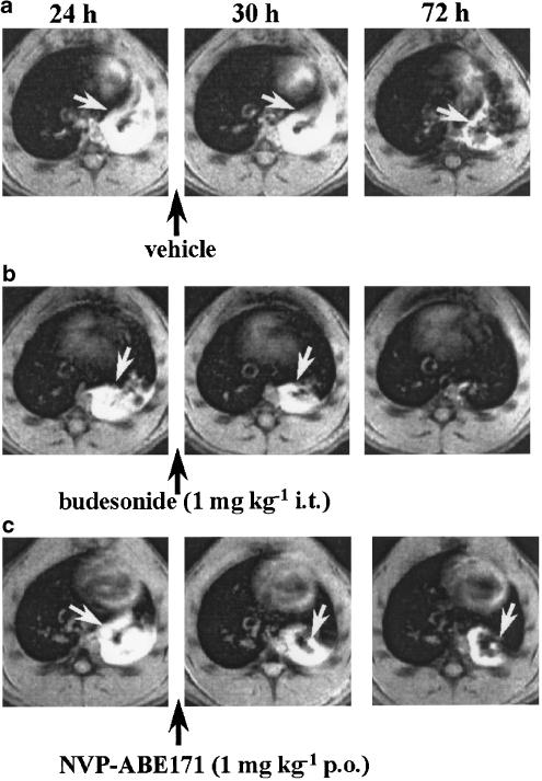
Figure 1.
Transverse sections through the thorax of BN rats acquired at different time points after challenge with OVA. Images correspond to approximately the same anatomical location in each animal. For each animal, the areas corresponding to oedematous signals (indicated by the white arrows) were assessed on 25 transverse sections analogous to those shown here and covering the chest. (a) NVP-ABE171 vehicle (2 ml kg−1 p.o.), (b) budesonide (1 mg kg−1 i.t.), or (c) NVP-ABE171 (1 mg kg−1 p.o.) was administered immediately after the 24 h MRI acquisition (indicated by the black arrows). MRI images were acquired at 24, 30 and 72 h after challenge with OVA (0.3 mg kg−1 i.t., time 0). Neither respiratory nor cardiac triggering was used, and the animals respired spontaneously during image acquisition.

