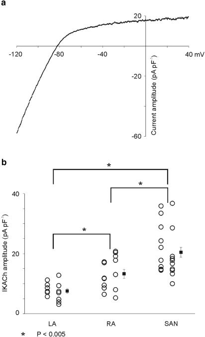
Figure 6.
Properties of IKACh in SA node myocytes. (a) The I–V relation of IKACh in an SA node myocyte. Comparison with Figure 2g shows that this I–V relation is very similar to that of atrial myocytes. (b) Comparison of data from individual myocytes (circles) plus mean±s.e.m. data (filled squares) for maximal IKACh current density at −50 mV in SA node myocytes with data from left and right atrial myocytes. IKACh current density is significantly larger in SA node myocytes than both left and right atrial myocytes. Also noteworthy is the degree of cell-to-cell variability of IKACh current density within both atria and within the SA node.

