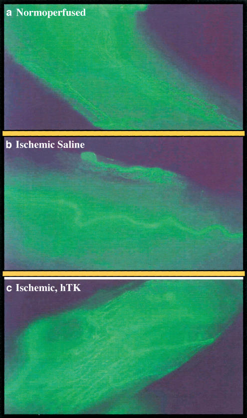
Figure 2.
Microphotographs showing the perineural microvasculature of sciatic nerves 21 days after induction of limb ischaemia produced in anaesthetized rats by surgical excision of left femoral artery. Contralateral normoperfused nerve microvasculature was counted for control. Animals (six per group) were injected with 50 μl sterile saline or 5 × l09 p.f.u. (in 50 μl saline) adenovirus containing the hTK gene or luciferase gene around the ischaemic sciatic nerve. At 21 days after induction of ischaemia, the animals were killed after sciatic nerve harvesting. Perineural microvasculature was visualized by in situ fluorescent staining using the endothelial cell-specific marker BS-1 lectin conjugated to FITC (Vector Laboratories, Burlingame, CA, U.S.A.). Images were captured using a Nikon Diaphot fluorescence microscope and an Optronics digital camera. Panel a shows the normal vascularization (in green) of normo-perfused nerve. Panel b shows rarefaction of vasa -nevorum network 21 days after ischaemia in saline-treated rat. Panel c shows the increased vascularity of sciatic nerve that was submitted to ischaemic insult and 3 days after receiving hTK gene transfer.

