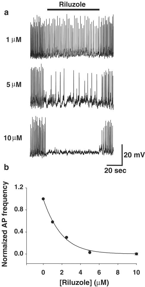
Figure 2.
Effect of riluzole on spontaneous action potentials in GH3 cells. (a) Traces show perforated patch recordings of spontaneous action potentials in the whole-cell current-clamp configuration. Spontaneous action potentials were inhibited in a dose-dependent fashion by riluzole. At 1 and 5 μM, riluzole reduced the frequency and amplitude of action potentials, whereas 10 μM riluzole abolished action potentials. See also Figure 5. (b) Plot of average action potential frequency (normalized to control frequency) vs riluzole concentration showing dose-dependent inhibition of action potentials.

